A. Brünott, E. Auriemma en A.B.M. Rijkhuizen
Hoofdafdeling Gezondheidszorg Paard, Faculteit der Diergeneeskunde, Universiteit Utrecht
e-mail: anne@brunott.biz
Summary
Objective: Description of the signalment, history, clinical signs and radiological findings of a horse with an acute severe lameness. Radiographs, ultrasonographic, CT and MRI images were taken to visualize the exact lesion.
Material and methods
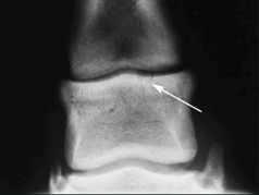
Based upon the history, clinical examination and diagnostic anaesthesia the region of pain was suspected to be the palmar aspect of the pastern joint region. After radiographic and ultrasonographic examination, CT and MRI were performed. A desmitis of the straight distal sesamoidean ligament and avulsion fragments from the proximal eminence of the middle phalanx (middle scutum) were diagnosed.
Results
The horse was treated conservatively with oral administration of non-steroidal anti-inflammatory drugs and submitted to a controlled exercise program. Within two weeks significant improvement was evident. After three months the horse had improved both clinically and on the views obtained by ultrasonography. A year later the horse was sound and had returned to his previous athletic level.


